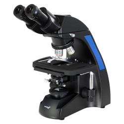
Digital inverted biological microscope MAGUS Bio VD300

MAGUS
Price: €2,785.67
Tax included
Availability Shipping in 3/7 days.
Code SGLV83012 Brand MAGUS EAN 5905555018096

The MAGUS Bio V300 is an inverted-design biological microscope for studying samples in laboratory glassware up to 70 mm high and with a bottom thickness of up to 1.2 mm. Specimens may include cell colonies, tissue cultures, biological fluids, etc. In addition, samples do not need to be stained to facilitate examination. Observations are made using transmitted light. Microscopy techniques: brightfield and phase contrast. This microscope is suitable for routine work in laboratories and research centers or for teaching.

Digital camera

Video optical tube with a SONY Exmor 8.3 MP digital sensor. The sensor uses back-illumination technology to improve light sensitivity and produce bright, sharp images even in low-light conditions. The camera is used for brightfield and darkfield microscopy with 4x, 10x, and 20x magnification objectives. It can also be used with stereo microscopes. A high-speed data transfer USB 3.0 port (5 Gbps) is used to connect to a computer and display images on the screen.

The MAGUS CDF30 camera is installed on the trinocular head or the optical tube of a microscope. The maximum field of view without optical defects can be achieved using an adapter with magnification from 0,65x to 0,9x. In addition to the adapter, an adapter with the same diameter as the tube is required to install the camera on the optical tube.

The image is displayed on a computer screen at a maximum resolution of 3840x2160 px. The frame rate is 45 fps. At a lower resolution (1920x1080 px), the frame rate increases to 70 fps. Transitions between frames are almost imperceptible, with no delays, making the camera suitable for studying and presenting moving objects. Photos and videos can be recorded and saved in various formats.

Optics

This microscope is equipped with a classic trinocular head, whose vertical tube is used to accommodate a digital camera. The camera is not included in the package. The beam splitter along the optical path has a 50/50 ratio.
The four included objectives, or other objectives purchased separately, can be mounted on the revolving nosepiece. All included objectives are designed for use with laboratory glassware and have a long working distance. Specifically, 3 plan achromatic objectives for brightfield observations and 1 plan achromatic objective for phase-contrast microscopy are included.

Illumination

The light source for transmitted-light observations is a 9 W LED. The high intensity of the LED provides sufficient illumination of the field of view for working with all the objectives and each of the available microscopy techniques. Adjusting the brightness does not cause any change in color temperature. The LED service life is approximately 50000 hours, with no need to replace the bulb.
The condenser has a slot for a phase-contrast slider (included). The phase-contrast rings can be centered. Inserting the slider saves time when switching from one microscopy technique to another.

Stage and focusing mechanism

Fixed mechanical stage. It is equipped with a plate holder (three holders for different sizes of glassware are included). The plates can be moved along the X and Y axes using a mechanical stage insert. The coaxial coarse and fine focusing knobs allow the image to be focused quickly and accurately. The coarse focus (coarse adjustment knob) has a locking mechanism, and the tension of the coarse adjustment knob can be adjusted.

Accessories

The range of optional accessories includes objectives, eyepieces, digital cameras, and calibration slides.

Key features of the microscope:

  • Brightfield and phase-contrast microscopy using transmitted light; study of samples in laboratory glassware
  • Digital camera installed in the trinocular tube
  • 9 W LED illuminator for transmitted-light observations, with adjustable brightness and a long service life
  • Fixed stage with movement along two axes; 3 plate holders of different sizes included
  • Optional accessories to enhance microscope performance

Key features of the camera:

  • Back-illuminated, color SONY Exmor CMOS sensor with high light sensitivity
  • Darkfield and brightfield operation using 4x, 10x, and 20x objectives
  • Connection to a computer via a USB 3.0 interface with a data transfer rate of 5 Gbps
  • Image displayed on a monitor at 4K Ultra High 3840x2160 px (45 fps) or Full HD 1920x1080 px (70 fps) resolution
  • Image-processing drivers and software included
  • Installation on the trinocular tube or optical tube of a microscope

The package contains:

  • MAGUS CDF30 digital camera (digital camera, USB cable, installation CD with drivers and software, user manual, and warranty certificate)
  • Stand with built-in power supply, transmitted-light source, focusing mechanism, stage, condenser with slot for a phase-contrast slider, and revolving nosepiece
  • Trinocular head
  • Infinity plan achromatic objective: PL 10x/0.25 WD 4.3 mm
  • Infinity plan achromatic objective: PL L20х/0.40 WD 8.0 mm
  • Infinity plan achromatic objective: PL L40х/0.60 WD: 3.5 mm
  • Infinity plan achromatic objective: PHP2 10x/0.25 phase contrast WD 4.3 mm
  • 10x/22 mm eyepiece with long eye relief (2 pcs.)
  • Eyecup (2 pcs.)
  • Centering telescope
  • Phase-contrast slider with centering phase-contrast rings
  • Mechanical stage for moving specimens
  • Plate holder (3 pcs.)
  • C-mount adapter
  • Color filter
  • AC power cable
  • Dust cover
  • User manual and warranty certificate

Available on request:

  • 10x/22 mm eyepiece with scale
  • 12,5x/14 mm eyepiece (2 pcs.)
  • 15x/15 mm eyepiece (2 pcs.)
  • 20x/12 mm eyepiece (2 pcs.)
  • 25x/9 mm eyepiece (2 pcs.)
  • Infinity-corrected plan achromatic objective: PL 4x/0.10 WD 21 mm
  • Infinity-corrected plan achromatic objective: PHP2 20x/0.40 phase contrast WD 8.0 mm
  • Infinity-corrected plan achromatic objective: PHP2 40x/0.60 phase contrast WD 3.5 mm
  • Calibration slide
  • Monitor and digital camera with HDMI interface
BrandMAGUS
Warranty5 years
EAN5905555018096
Package size (LxWxH)64x42x67 cm
Shipping weight18.6 kg
=== Delimiter ===Microscope specifications
Typebiological, digital, light/optical
Microscope head typetrinocular
HeadSiedentopf
Head inclination angle45°
Magnification, x100 — 400
Magnification rangefrom 200x to 800x
Magnification, x (optional)40–500/600/800/1000
Eyepiece tube diameter, mm30
Eyepieces10x/22 mm, eye relief: 10 mm (*optional: 10x/22 mm with reticle, 12,5x/14; 15x/15; 20x/12; 25x/9)
Objectivesinfinity-corrected plan achromatic: PL 10x/0.25; PL 20x/0.40; PL 40x/0.60; PHP2 10x/0.25 phase; parfocal distance: 45 mm (*optional: PL 4x/0.10; PHP2 20x/0.40 phase; PHP2 40x/0.60 phase)
Nosepiecefor 4 objectives
Working distance, mm21.0 (4x); 4.3 (10x); 8.0 (20x); 3.5 (40x)
Interpupillary distance, mm48 — 75
Stage movement range, mm77/112
Stage featuresfixed, with mechanical stage for moving the specimen; plate holders: 29x77 mm, Ø90 mm; 34x77,5 mm, Ø68,5 mm; 57x82 mm, Ø60 mm
CondenserN.A.0.6, working distance: 70 mm; with adjustable aperture diaphragm and a slot for a phase contrast slider
Diaphragmadjustable aperture
Focusingcoaxial, coarse focusing (with tension adjustment knob and locking knob) and fine focusing (micrometer screw at 0.002 mm)
Bodysturdy aluminum
IlluminationLED
Brightness adjustmentyes
Power supply220±22 V, 50 Hz, AC mains
Light source type9 W
Optical filtersyes
Operating temperature range, °C5 — 35
Special featuresphase contrast slider for 10x objectives with a centering phase contrast ring
User levelprofessionals, experienced users
Assembly and installation difficulty levelcomplicated
Applicationlaboratory/medical
Illumination positionupper (transmitted light)
Research methodbrightfield, phase contrast microscopy
Set with case/cover/bagdust cover
=== Delimiter ===Digital camera
SensorSONY Exmor CMOS
Color/monochromecolor
Megapixels8.3
Maximum resolution, px3840x2160
Sensor size1/1.2'' (11,14x6,26 mm)
Pixel size, µm2.9x2.9
Light sensitivity5970 mV/lux/s
Signal-to-noise ratio0.13 mV at 1/30 s
Exposure time0.02 ms – 15 s
Video recordingyes
Video recordingyes
Frame rate, fps@resolution45@3840x2160, 70@1920x1080
Installation locationTrinocular tube, optical tube instead of eyepiece
Image format*.jpg, *.bmp, *.png, *.tif
Video formatrecording: *.wmv, *.avi, *.h264 (Windows 8 and later), *h265 (Windows 10 and later)
Spectral range, nm380-650 (integrated IR filter)
Shutter typeERS (electronic rolling shutter)
White balanceautomatic, manual
Exposure controlautomatic, manual
Software functionalityexposure control, image size, brightness
SoftwareMAGUS View
Output5 Gb/s, USB 3.0
System requirementsWindows 8/10/11 (32-bit and 64-bit), Mac OS X, Linux, up to 2.8 GHz Intel Core 2 or later, minimum 2 GB RAM, USB 3.0 port, CD-ROM, 17" or larger
Mount typeC-mount
Camera power supplyDC, 5 V, from the computer's USB port; 12 V adapter, 3 A for the Peltier cell
Camera operating temperature range, °C-10 — 50
Operating humidity range, %30 — 80

Digital inverted biological microscope MAGUS Bio VD300

MAGUS
Price: €2,785.67
Tax included
Noleggialo a €56.54 + IVA / mese per 60 mesi con Maggiori informazioni ›
Availability Shipping in 3/7 days.
Code SGLV83012 Brand MAGUS EAN 5905555018096
Share on
Comments (0)