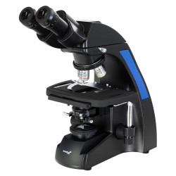
Digital inverted biological microscope MAGUS Bio VD300 LCD
- Over 90,000 satisfied customers since 2020
- Secure payments: credit card, PayPal and bank transfer
- Expedited shipping: Poste, BRT, FedEx
- Customer support Whatsapp +39 371 43 61 201
The MAGUS Bio VD300 LCD is an inverted-design biological microscope for studying samples in laboratory glassware up to 70 mm high and with a bottom thickness of up to 1.2 mm. Samples may include cell colonies, tissue cultures, biological fluids, etc. Samples do not need to be stained to facilitate examination. Observations are made using transmitted light. Microscopy techniques: brightfield and phase contrast. This microscope is suitable for routine work in laboratories and research centers, or for teaching.
Digital camera
The MAGUS CHD30 digital camera is equipped with a 2 MP sensor and produces realistic Full HD images at a resolution of 1920x1080 px. The camera has high light sensitivity and an autofocus function, and is suitable for fluorescence observations.
The camera uses an HDMI interface to connect directly to a TV monitor or projector. This allows the camera to operate independently, without a PC connection. The HDMI interface enables fast, stable transfer from the camera to the external screen. It can be connected to a PC via Wi-Fi.
Video is recorded at 60 fps or 25 fps, depending on the video output interface.
The camera combines a high FPS rate with high HDMI bandwidth. Videos are therefore bright, with no freezing or dropped frames. At maximum resolution, the image is highly detailed, moving objects are visible without errors, and motion is displayed without delays.
Monitor
MAGUS MCD monitors work in combination with MAGUS CHD cameras to form a complete imaging system for MAGUS microscopes.
The camera connects via its HDMI interface. The monitor displays images in real time. Its IPS LCD display has a wide viewing angle, high brightness, and good contrast. Even when viewing the screen from a wide angle, the image is not distorted. The screen size is 13.3''.
The monitor can be placed on a horizontal surface using a folding stand or mounted directly on a camera or tripod.
Optics
This microscope is equipped with a classic trinocular head, whose vertical tube is used to accommodate a digital camera. The camera is not included in the package. The beam splitter along the optical path has a 50/50 ratio.
Four included objectives or others purchased separately can be mounted on the revolving nosepiece. All included objectives are designed for use with laboratory glassware and have a long working distance. Specifically, the package includes 3 plan achromatic objectives for brightfield observations and 1 plan achromatic objective for phase-contrast microscopy.
Illumination
The light source for transmitted-light observations is a 9 W LED. The LED's high intensity provides sufficient illumination of the field of view for working with all objectives and each of the available microscopy techniques. Adjusting the brightness does not change the color temperature. The LED lifespan is approximately 50000 hours, with no need to replace the bulb.
The condenser has an available slot for a phase-contrast slider (included). The phase-contrast rings can be centered. Inserting the slider saves time when switching from one microscopy technique to another.
Stage and focusing mechanism
Fixed mechanical stage. It is equipped with a plate holder (three holders for glassware of different sizes are included). The plates can be moved along the X and Y axes using a mechanical stage insert. The focusing mechanism with coaxial coarse and fine adjustment knobs enables rapid and accurate image focusing. The coarse focusing knob is equipped with a locking mechanism, and its tension can be adjusted.
Accessories
The optional accessories range includes objectives, eyepieces, digital cameras, and calibration slides.
Key microscope features:
- Brightfield and phase-contrast microscopy using transmitted light; sample observation in laboratory glassware
- Digital camera installed in the trinocular tube
- 9 W LED illuminator for transmitted-light observations, with adjustable brightness and a long lifespan
- Fixed stage with movement along two axes; 3 plate holders of different sizes included
- Optional accessories to improve microscope performance
Key camera features:
- The camera operates independently, without a PC connection, via the HDMI interface. It can be connected to a PC via the USB 2.0 interface
- Autofocus in HDMI mode makes it easier to adjust image sharpness
- The camera has a resolution of 1920x1080 pixels, an ideal choice for displaying images on a Full HD monitor
- 60 fps for observing moving objects, recording video, and moving the specimen without delays or jerks
- 25 fps via Wi-Fi, sufficient for adjusting focus and processing images using the software
- SONY color CMOS sensor, low noise, and high light sensitivity
- Software for recording and editing photos and videos, external display functions, linear and angular measurements
The package includes:
- MAGUS CHD30 digital camera (digital camera, HDMI cable (1.5 m), mouse with USB, 32 GB SD memory card, USB Wi-Fi adapter, 12 V/1 A AC power adapter (Euro), installation CD with drivers and software, user manual, and warranty certificate)
- MAGUS MCD20 LCD monitor
- Stand with integrated power supply, transmitted-light source, focusing mechanism, stage, condenser with slot for phase-contrast slider, and revolving nosepiece
- Trinocular head
- Infinity plan achromatic objective: PL 10x/0.25 WD 4.3 mm
- Infinity plan achromatic objective: PL L20x/0.40 WD 8.0 mm
- Infinity plan achromatic objective: PL L40x/0.60 WD: 3.5 mm
- Infinity plan achromatic objective: PHP2 10x/0.25 phase contrast WD 4.3 mm
- 10x/22 mm eyepiece with long eye relief (2 pcs.)
- Eyecup (2 pcs.)
- Centering telescope
- Phase-contrast slider with centering phase-contrast rings
- Mechanical stage for moving samples
- Plate holder (3 pcs.)
- C-mount adapter
- Color filter
- AC power cable
- Dust cover
- User manual and warranty certificate
Available on request:
- 10x/22 mm eyepiece with graduated scale
- 12,5x/14 mm (2 pcs.) eyepiece
- 15x/15 mm (2 pcs.) eyepiece
- 20x/12 mm (2 pcs.) eyepiece
- 25x/9 mm (2 pcs.) eyepiece
- Infinity-corrected plan achromatic objective: PL 4x/0.10 WD 21 mm
- Infinity-corrected plan achromatic objective: PHP2 20x/0.40 phase contrast WD 8.0 mm
- Infinity-corrected plan achromatic objective: PHP2 40x/0.60 phase contrast WD 3.5 mm
- Calibration slide
| Brand | MAGUS |
| Warranty | 5 years |
| EAN | 5905555018102 |
| Package size (LxWxH) | 64x42x67 cm |
| Shipping weight | 20.75 kg |
| === Delimiter === | Microscope specifications |
| Type | biological, digital, light/optical |
| Microscope head type | digital screen/PC monitor, trinocular |
| Head | Siedentopf |
| Microscope head inclination angle | 45° |
| Magnification, x | 100 — 400 |
| Magnification range | from 200x to 800x |
| Magnification, x (optional) | 40–500/600/800/1000 |
| Eyepiece tube diameter, mm | 30 |
| Eyepieces | 10х/22 mm, eye relief: 10 mm (*optional: 10x/22 mm with graduated scale, 12,5x/14; 15x/15; 20x/12; 25x/9) |
| Objectives | infinity-corrected plan achromatic: PL 10x/0.25; PL 20x/0.40; PL 40x/0.60; PHP2 10x/0.25 phase; parfocal distance: 45 mm (*optional: PL 4x/0.10; PHP2 20x/0.40 phase; PHP2 40x/0.60 phase) |
| Objective turret | for 4 objectives |
| Working distance, mm | 21.0 (4x); 4.3 (10x); 8.0 (20х); 3.5 (40x) |
| Interpupillary distance, mm | 48 — 75 |
| Stage travel range, mm | 77/112 |
| Stage features | fixed, with mechanical specimen translation; plate holders: 29x77 mm, Ø90 mm; 34x77,5 mm, Ø68,5 mm; 57x82 mm, Ø60 mm |
| Condenser | N.A.0.6, working distance: 70 mm; with adjustable aperture diaphragm and a slot for a phase contrast slider |
| Diaphragm | adjustable aperture |
| Focus | coaxial, coarse focusing (with tension adjustment knob and focusing lock knob) and fine focusing (micrometer screw of 0.002 mm) |
| Body | CNC-machined aluminum alloy |
| Illumination | LED |
| Brightness adjustment | yes |
| Power supply | 220±22 V, 50 Hz, AC 110–220 V, DC 5–12 V/1 A (Type C), AC mains |
| Light source type | 9 W |
| Optical filters | yes |
| Operating temperature range, °C | 5 — 35 |
| Special features | phase contrast slider for 10x objectives with a centering phase contrast ring |
| Ability to connect additional equipment | Wi-Fi adapter (USB), mouse with USB |
| User level | professionals, experienced users |
| Assembly and installation difficulty level | complicated |
| Application | laboratory/medical |
| Illumination position | upper (transmitted light) |
| Research method | brightfield, phase contrast microscopy |
| Set with case/cover/bag | dust cover |
| === Delimiter === | Digital camera |
| Sensor | SONY CMOS |
| Color/monochrome | color |
| Megapixels | 2 |
| Maximum resolution, px | 1920x1080 |
| Sensor size | 1/1.9" (7,20x4,05 mm) |
| Pixel size, µm | 3.75x3.75 |
| Autofocus | yes |
| Interface connectors | HDMI 1.4, Wi-Fi |
| Memory card | SD up to 32 GB |
| Wi-Fi settings | 802.11n, 150 Mbps, 20 dBm |
| Light sensitivity | 1120 mV at 1/30 s |
| Signal-to-noise ratio | 0.15 mV at 1/30 s |
| Exposure time | 0.06 ms – 918 ms |
| Video recording | yes |
| Frame rate, fps@resolution | 25@1920x1080 (Wi-Fi), 60@1920x1080 (HDMI) |
| Installation location | trinocular tube, optical tube instead of eyepiece |
| Image format | *.jpg |
| Video format | *.asf |
| Spectral range, nm | 380-650 (built-in IR filter) |
| Shutter type | ERS (electronic rolling shutter) |
| Software | HDMI: built-in; USB: MAGUS View |
| System requirements | Windows 8/10/11 (32-bit and 64-bit), Mac OS X, Linux, up to 2.8 GHz Intel Core 2 or later, minimum 4 GB RAM, USB 2.0 port, RJ45, 19" or larger display (with USB connection) |
| Mount type | C-mount |
| Camera power supply | DC adapter 12 V, 1 A |
| DC power adapter specifications | input: AC voltage 100–240 V, 50/60 Hz, output: DC voltage 12 V/1 A |
| Camera operating temperature range, °C | -10 — 50 |
| Operating humidity range, % | 30 — 80 |
| === Delimiter === | Monitor |
| Matrix type | IPS |
| Display diagonal, inches | 13.3 |
| Display resolution, px | 1920x1080 (Full HD) |
| Aspect ratio | 16:9 |
| Brightness, cd/m² | 400 |
| Number of colors displayed | 16.7 m |
| Contrast | 1000:1 |
| Horizontal/vertical viewing angle, ° | 178/178 |
| Screen viewing area dimensions (W×H), mm | 295x165 |
| Pixel pitch (W×H), mm | 0,154x0,154 |
| Optical source frequency, Hz | 60 |
| Matrix-type backlight | LED |
| LED backlight lifespan, h | 50000 |
| Interface | HDMI |
| Power consumption, W | 12 (max.) |
Digital inverted biological microscope MAGUS Bio VD300 LCD
Locazione Operativa
Accedi alle migliori tecnologie senza immobilizzare capitali.
| Durata | Canone mensile | Totale |
|---|---|---|
| 24 mesi | €126.38 + IVA | €3,033.12 + IVA |
| 30 mesi | €100.66 + IVA | €3,019.80 + IVA |
| 36 mesi | €87.75 + IVA | €3,159.00 + IVA |
| 48 mesi | €66.74 + IVA | €3,203.52 + IVA |
| 60 mesi | €55.42 + IVA | €3,325.20 + IVA |
* Importi al netto di IVA. Canoni calcolati sul prezzo del prodotto ex-IVA.
📋 Condizioni contrattuali (applicate da Grenke, non in carrello)
- Durata: da 24 a 60 mesi
- Importo noleggiabile: da €500 + IVA
- Pagamento: trimestrale anticipato tramite RID
- Assicurazione: 1,95% annuo sul valore del bene (fatturata direttamente da Grenke)
- Spese di incasso: € 0
-
Spese amministrative:
Fino a €12.500: €75 + IVA €12.501 – €25.000: €90 + IVA €25.001 – €100.000: €100 + IVA Oltre €100.000: €200 + IVA
- Riscatto a scadenza: Restituzione del bene oppure riscatto al 3% del valore iniziale del contratto
- Requisiti: Titolari di Partita IVA con almeno 2 anni di attività, con indirizzo in Italia
Le spese amministrative e assicurative sono gestite direttamente da Grenke e non vengono addebitate dal negozio. I canoni indicati sono stimati sul prezzo del prodotto e potranno essere confermati da Grenke in fase di istruttoria.
- Over 90,000 satisfied customers since 2020
- Secure payments: credit card, PayPal and bank transfer
- Fast shipping: Post and BRT
- Customer support Whatsapp +39 371 43 61 201
Your review appreciation cannot be sent
Report comment
Report sent
Your report cannot be sent
Write your review
Review sent
Your review cannot be sent